This hormone is produced in a section of the brain known as the hypothelamis
Like all hormones it flows through the bloodstream
Its target is the kidney.
The effect of this hormone is to control the quantity of water in blood as it is important that the tissue fuid is isotonic in every cell.
ADH actually targets the collecting duct. The effect of this is that it allows more water to come out of the collecting duct.
It is possible to increase the amount of water going into the blood through the collecting duct by applying ADH. It makes the collecting duct wall more porus therefore more water can escape from the collecting duct and flow into the bloodstream.
The consequences of this ADH excretion is that the urine would be more concentrated and it would have a lower volume.
We have blood coming into the kidney. It is placed under high pressure.
The dissolved contents in the blood are forced into the bowman's capule's tube, and would become known as the glomerula filtrate.
This glomerula filtrate contained salts, water, glucose and urea.
When the filtration occurs, it filters out too much water.
The filtrate passes along the tube and once it reaches the collecting duct, water is removed from the filtrate.
that water is returned back to blood vessels therefore the water has been selected and has been reabsorped into the blood. We get the word, selective reabsorption.
Selective reabsorption takes place in the collecting duct.
Nephron is the tubular structure in the kidney. This is the structure which carries out the filtration of our blood and ultimately results in two things
Filtered blood
Urine
Thr urine that comes out of the plevic region is composed largely of water, salts, and molecule known as urea (nitrogen waste).
The process of filtration, known as ultra filtration, filtration of molecules, starts in the bowman's capsule.
FIltration of blood begins when the blood arrives in the kidney, in the nephron, in the vessel known as the aferet arteriole. The blood here is under high pressure
The blood vessel coming out of the bowman's capsule is known as the efferent arteriole. The diameter of this blood vessel is much smaller than the afferent arteriole.
The blood develops a high pressure as it flows from a wide blood vessel (afferent arteriole) to a narrow blood vessel (efferent arteriole), the blood pressure increasing in the glomerulis.
The high pressure forces the liquid within the blood (plasma) and is forced into the space within the insidde of the bowman's capsule.
The plasma then becomes known as the glumerula filtrate.
The branch of the aorta taking blood into the kidney is known as the renal artery
Th kidney filters the blood and the contents which are removed and excreted from the blood are called urine, which comes down the structure known as the ureter, and is then collected in the bladder for release.
The filtered blood exits through the renal vein and would return to the vena cava .
If we slice through the kidney to show the internal structure, we see different colored regions
The outer lighter region is known as the cortex, and the inner darker region is known as the medulla and the innermost lightest region is known as the pelvic region.
The urine is collected in the pelvic region from where it drains down the ureter.
The reason for these different colors is because the kidney is made up of various tubular structures.
The tubular structure can be shown in here:
The tube structure is known as the nephron.
The first twisted section is known as the proximal convoluted tubule section.
The second twisted section is known as the distal convoluted tubule section.
There are millions of these nephrons (tubular structure) in the kidney.
two kidneys- a left one and a right one, each with its own blood supply, carrying out the process of excretion, filtration, and osmoregulation. From each kidney there is a tube that leads to the bladder, which is called a ureter.
ureter-carries urine from the kidney to the bladder.
Bladder- the urine is conducted to the outside of the body to be excreted down a structure called the urethra, which either travels down through the vagina or the penis.
We have cells in the body and ideally if the tissue fluid that surround these cells look like the ones in he diagram, this must be isotonic with the cytoplasm of these cells.
This means that the amount of water going outside and inside of the cell is equal which further means that the cell will maintain its shape, size and function.
The danger to the tissue is that the blood circulating to the tissue would be concentrated causing a hypertonic tissue fluid or it may be dilute causing a hypotonic tissue fluid.
Hypertonic would remove too much water and hypotonic would add too much water to the cell.
We want to keep the tissue fluid isotonic to the cell's cytoplasm.
This achieved by controlling the composition of blood.
Blood forms the tissue fluid. It is the role of the kidney to control the composition of blood
Blood which circulates through our kidney, excess water, excess salts can be removed and excreted through the uretus.
If the kidney can control the content of water and salts in the blood, the kidney can keep the blood and therefore the tissue fluid isotonic with the cell's cytoplasm, maintaining the function of the cell
photosynthesis-involves the leaf absorbing sunlight and while doing so it combines carbon dioxide and water to produce glucose and oxygen, a gas which is given off.
In this case, the gas which is given off, oxygen, is an example of excretion as its a waste molecule.
Excretion- the release of metabolic waste.
Respiration-glucose and oxygen are used, and through a series of enzyme reactions, the glucose molecules break down and the energy is used to form ATP, water, and carbon dioxide. In this process, the waste product is carbon dioxide and water. However, like the oxygen in the reaction above, Carbon dioxide is also given off, its a metabolic waste therefore its excretion.
Plants excrete oxygen when they photosynthesize and they excrete Carbon dioxide when they go through the process of respiration.
Staphlococcus aureus-bacteria which causes skin infection and some lung infection.
Those who are affected by this bacteria can be treated by an antibiotic known as methecilline.
This antibiotic kills the staphlococcus aureus.
The type of staphlococcus aureus which can be killed by the antibiotic are known as susceptible form (mssa)
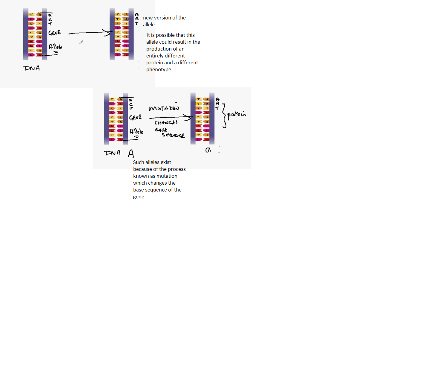
When a random mutation occurs within the genome of the staphlococcus aureus, the methecilline fails to kill it. This form is known as the resistant form of staphlococcus aureus. (mrsa)
The mutation has created genes within the bacteria has allowed it to break down the antibiotic.
Variations- differences we can see in the phenotypes of each individual.
It is possible to count/measure these differences and show them through graphs.
The phenotype depends on the genotype of each individual which is modified according to the environment.
Variation in population is a variation within all these individuals.
The difference in appearance within different species occur because all those individuals have different genotypes and they survive in different environments.
Phenotypic variation in a population or species is due to environmental variation. E.g. the home language you speak.
The parent phenotype is red petal crossed with red petal
The parent genotype- Rr x Rr
Both parents are heterozygous.
Meiosis takes place and the allele has to be separated, one into each type of gamete.
In this case, there is a 50% chance that the pollen grain is carrying a R allele and there's a 50% chance that the pollen grain is carrying an r allele.
Random fertilization occurs, as shown in the table, to be observed in the image.
The probabilities depend on which pollen grain (R or r) fertilizes with which ovule (R or r)
We're predicting the probabilities of offspring through monohybrid crosses.
Monohybrid crossing- crossing which consists of only one gene.
Let's consider the cross between a red petal plant and a white petal plant.
The genotype of the red petal plant is RR and the genotype for a white petal plant is rr.
This makes the R>r. In other words, red is dominant to white.
The next stage involves meiosis which includes the production of the pollen grains and the ovules.
The gamete will contain only one of the two alleles.
For instance, in the production of the pollen, 1/2 the gametes will have the R and the other half will have the other big R as its an equal chance.
In the production of the ovules we need to separate the alleles so that we only have 1 of the pair of alleles in each gamete. half of the ovules would have little r and the other half would have little r.
Oestrogen and Progesterone are both examples of hormones.
Hormones are produces in a structure called endocrine gland.
Hormone will travel through the blood from the endocrine g;and to its target tissue.
The hormone will have an effect on the target tissue. Some hormones such as oestrogen and progesterone will have multiple effects.
The ovary is the endocrine gland for oestrogen.
The oestrogen flows from the ovary through the bloodstream and into the lining of the uterus. The effects of this are:
In the first half of the cycle (14 days) the lining becomes increasingly thick. The oestrogen brings about the thickening of the wall of the endometrium.
oestrogen flows through our bloodstream to our brain. In the brain, it releases another hormone called liutenizing hormone (LH). The production of this hormone reaches its peak at about day 13 in the cycle. This causes the ovary to release an egg. The egg will come out of the ovary and will move into the oviduct where it is now possible for fertilization to occur.
Whilst this is happening, a circular structure within the ovary known as a folicle becomes larger and larger. The egg is inside the folicle. It is the cells around the folicle which produce oestrogen.
The folicle reaches its largest size at about day 13 when the liutenizing hormone causes the wall of the folicle to rupture to release the egg.
This process is known as ovulation.
After the completion of this process, the empty follicle has an altered function. It develops a yellow color called corpus liuteum.
This yellow body is known to produce progesterone. The ovary and the corpus luteum is the endocrine gland for progesterone.
The progestrone flows through the bloodstream to the lining of the uterus and maintains the thickness of it for the next (16-24) 8 days of the cycle. It prevents the lining from breaking down.
During this period of time, there is a possibility of the fertilized egg implanting into the wall of the uterus and developing into a pregnancy.
However, if this does not happen, the corpus luteum breaks down and the progesterone level falls leading to the breakdown in the lining of the uterus.
This concludes with the release of menstrual period.
Looking down a microscope, at a cell, we can see the nucleus, a spherical structure. However, the chromosomes would not be seen.
It is during the interphase where the DNA replication takes place.
The first sign of mitosis, we see when we look down a microscope, is when the nuclear membrane breaks down and the chromatids (pairs of chromosomes) become visible. This is a phase known as the prophase.
Now that the nucleus is gone, inside the cell there is a network of protein molecules known as the spindle, each made out of a fibre. These extend from one pole of the cell to the other. During late prophase, the pair of chromatids will move towards the spindle and will join onto one of the spindle/fibre at the centromere.
The next stage is known as the metaphase:
The pair of chromatids is attached to the spindle firbe by the centromere.
The characteristicof this stage is that the chromosomes are in the middle, arranged across the equator of the cell.
Anaphase:
The spindle/fibre shortens, pulling one chromatid upwards and the other downwards.
The pair of chromatids are moving apart to the poles. the chromatids are being separated.
Telophase:
The chromosomes are at opposite ends of the cell.
The nucleus begins to reform around the chromosomes at either end of a cell.
These will be the new nuclei of the new cells
In the telophase we see the formation of two nuclei
Cytokinesis:
In this process, the cell splits into two.
This is not a part of mitosis.
We see the nucleus that had reformed in the anaphase.
The cell begins to divide its cytoplasm in half
The membrane of the cell fuses across the equator to form the two cells
The two new cells will contain 1 chromosome just like the parental cells do.
In human cells we won't see one pair of chromosomes separating, but 23 separating at the same time.
The cell which will undergo the process of mitosis will have to copy its chromosomes.
The process of copying chromosomes is called DNA replication. In this process each chromosome undergoes a copying process to form an identical copy of itself, with the same genes.
These pairs of copies are held together through a bonding in the centre region called the centromere.
These pairs are referred to as a pair of chromotids
The process of DNA replication takes place in the nucleus while its still intact. We cannot see the process.
This is known as the interphase of the cell cycle. It is during this resting stage of the cycle during which the chromosomes are copied.
Mitosis is a type of cell division which causes growth which occurs via the increasing numbers of cells.
The number of chromosomes in a nucleus is known as a diploid number (abbreviated as 2n).
The diploid number in a human cell is 46. 2n = 46
for cats 2n = 38
In the process of mitosis a particular cell divides into two cells, each with a nucleus. When we look inside the nucleus of these cells, we find that each of these cells have a diploid nucleus.
These cells are identical:
They have the same number of chromosomes
Same set of chromosomes- both cells have identical chromosomes, to each other.
Sexual reproduction involves sexes which are identified as males and females.
Organisms, who reproduce sexually, produce cells known as gametes. This takes the form of a sperm cell in the male and an egg cell in the female.
The type of cell division that produces gametes are known as meiosis.
Meiosis have two effects. One of which is to half the number of adult chromosomes in a gamete cell.
The total number of chromosomes in an adult is 46 per cell whereas the total number of chromosomes in a gamete cell is 23.
Going from 46 to 23 is the process of cell division known as meiosis.
We have the process of fertilization in which the sperm cell fuses with the egg cell.
We find variation in the sexually producing population. This variation (differences) is broad. We find many differences in an individual of the sexually reproducing population.
Asexual:
No gametes
No sexes.
Process of cell division is known as mitosis in eucaryotic cells and binary fission in procaryotic cells which exist in bacteria.
In this process the number of chromosomes is maintained constantly. E.G. a cell with 20 chromosomes would divide to produce 20 chromosomes in 2 cells. These two cells would be identical.
Small amount of variation due to mutation. They are usually identical. This is known as a clone.
The greenhouse effect is brought about by pollution.
Molecules such as Carbon dioxide, water vapour, and methane are greenhouse gases.
The concentration of these gases reemit the infrared light back to earth instead of allowing it to escape into space. This raises the average global temperature. This is known as Global warming.
The consequences of global warming are the melting of ice caps in the polar regions, resulting in the rise of sea levels. This would change the ocean currents. It would also change the direction of the wind.
This would result in climate change.
Raising the average global temperature would change the distribution of the world's biomes (major vegetation ecosystems). Due to this deserts would expand
Short wavelengthed UV light is passed into the atmosphere.
50% of this light is reflected back into earth.
The other 50% is absorbed by the earth's surface. The UV light is converted and transferred as infrared.
The infrared is emmitted back outwards-some of this is lost in space in the form of heat.
Major examples of the greenhouse gases are water vapour, methane, carbon dioxide.
Infrared waves hit these gases. the gases then reemits these waves in all directions, including downwards. This raises the temperature slightly.
Enhanced Greenhouse Effect consists of increases amounts of Carbon Dioxide and methane. This causes more radiation of infrared waves causing the temperature to rise. This then leads to climate change.
Chloroflurocarbons are well known for the affect on the ozone layer.
This catalizes the breakdown of O (to the power of 3) to O (to the power of 2).
O3 is much better at absorbing UV light therefore by releasing CFCs we are removing the protection of the ozone layer.
The Pollution is caused by sulphur dioxide and carbon monoxide.
Sulphur dioxide is added to the atnosphere through the combustion of fossil fuels.
The combustion of these fossil fuels results in harmful gases released into the atmosphere.
Sukphur dioxide is also released through fuel engines in cars.
In the atnosphere, Sulphur dioxide combines with water vapour and forms sulphuric acid.
The sulphuric acid is then stored in the clouds which condense to form rainfall. This type of rain is called acid rain.
Acid rain burns trees and plants due to the direct effect of the sulphuric acid on the surface of the leaves.
Sukphuric acid also affects the plants by leaching calcium and magnesium ions in the soil. This results in the plant not being able to grow.
The acid rain precipitation forms streams which will join the lakes. This will reduce the pH and release aluminium ions. This will kill the fish as aluminium will thicken the mucus that lines the gills of the fish. This reduces oxygen supply to the fish.
Carbon Monoxide is also produced when fossil fuels are burned with insufficient oxygen. Incomplete combustion.
Carbon monoxide combines with the haemoglobin in the red blood cells and forms a molecule known as carbaminohaemoglobin.
This blocks haemoglobin from carrying oxygen. This decreases the amount of oxygen being supplied to pther parts of the body.
We have the lungs on top and then the heart followed by the liver and the kidney.
Blood which is travelling from the right ventricle travels to the lung therefore it's a polmonary artery.
The blood returns to the heart through the polmonary veins and enters the left atrium.
Blood leaves the left atrium through the aorta and is transported to the body. The aorta branches to the liver. The smaller blood vessel branches out of the aorta known as the hepatic artery.
A branch of the aorta also travels to the kidney. The blood vessel is known as the reinal artery. Blood returns to the heart in a blood vessels travelling up from the kidney. Blood flows into the heart through the veena cava.
Blood passes from the liver (hepatic vein) through the capillaries and then back into the heart via the vena cava.
The artery is carrying high blood pressure whereas the vein is carrying is carrying low blood pressure.
The capillary is the site for exchange.
The capillary walls are very thin, only one cell thick, easy for diffusion of gases.
The wall of the artery is thick and contains muscle. The lumein carries the blood is narrow. The muscle contracts, it maintains the blood pressure all the way to the organ.
Veins are collecting blood and returning them to the heart. The walls are thin compared to the artery. Large lumein which has low resistance, blood can flow through easily. They have valves to stop backflow.
All arteries take blood away from the heart. There blood is under high pressure. Blood is being delivered to another organ through arteries.
All veins take blood to the heart. Veins contain low pressured blood. Blood in veins fro an organ.
Between the artery and vein, the blood vessels travel through the organ. This is where we have exchange where the contents of the cell is exchanged with the blood's contents.
The blood vessels in an organ are called capillary.
Substances go into and out of the cell. e.g. carbon dioxide and oxygen. Nothing goes into the vein or the artery, nor does it go out. The place for exchange is the capillary.
If blood is going into the heart, the blood vessel is called a vein. If blood is going out of the heart that kind of blood vessel in called an artery.
The technical word for lung is polmonary. We have the polmonary artery which takes the blood from the heart to the lung.
The polmonary vein takes the blood from the lung to the heart.
The major artery in the body is the aorta which takes blood from the heart to around the body.
The major vein in the body is Vena cava.
These two are our major blood vessels.
The heart has two small chambers: the left atrium and the right atrium.
The large chambers are known as the keft and rught ventricles.
What separeates the right and lift side of the heart is the septum.
The valves on the left side are called the bicuspid valves which open and close, allowing blood to flow through.
The valves in the right side are called the tricuspid valves.
The valves beneath the aorta and polmonary artery are called the semilunar valves.
The bicuspid valves: When the pressure is high above the valves, the blood forces down through the valves, forcing the cusps open. The blood then flows to a region of low pressure.
When the ventricle contracts we get high pressure below the bicuspid/tricuspid valves, causing it to close. This makes a sound 'lub' or 'lup'
The semilunar valves react in the same way. When the high pressure is below the valves, the blood is forced upwards into the arteries, opening the cusps. However, when the the heart relaxes the blood flows back down, closing the semi lunar valves. This is the second sound known as 'dub'.
This overall, gives a lub dub sound. 'Lub' is the sound caused by the closure of atrial ventricular valves whereas 'dub' is caused by the closure of the semilunar valves.
Platelets are produced in bone marrows and they are fragments of cells.
When we have a wound, the platelets in the blood would be exposed to the air and therefore they release chemicals.
In the blood there is a protein called fibrinogen and this is soluble. However from the chemical in the platelets this turns into firbin.
This results in us getting a metrix, a netwrok of fibrin molecules on the wound and onto this solidifies red blood cells. This will dry over to form a scab beneath which the cells will begin to repair the wound.
The clotting which occurs right below the wound stops blood loss.
White blood cells will attack any pathogens present in the wound or underneath the wound.
Pathogen is a disease causing organism e.g. bacteria.
Lymphocite is a type of white blood cell. It had a large nucleus.
Each bacteria has a specific type of lymphocyte.
When the bacteria and the lymphocyte come together, the White blood cell divides into two clones.
One clone is called memory cells while the other clone releases antibodies and is known as plasma cell.
These plasma cells release from the cell into the bloodstream, protein molecules which are our antibodies.
The antibody attaches to the bacterial cell an act as a label that will atract phagocytes or the antibody attatches and causes bacterial lysis, the cell bursts.
The antibody causes the bacterial cells to stick together. They cause many bacteria to stick to gether. This is called glutination. This results in a phagocyte engulfing many bacteria at the same time.
The chances of the bacteria meeting the memory cells is greater than meeting the one original cell. This results in us reacting faster with more antibodies. This is called the secondary response.
White blood cells have lobed nucleus. It defends us against infections. It can detect the bacteria because the bacteria gives off chemicals. Phagocytes is a type of white blood cell.
'phago' means feeding and 'cites' mean cells.
When the phagocytes sense the chemicals from the bacteria, they extend themselves around the bacteria. These extensions are called psaedopodia. 'Psaedo' means false and 'podia' means food.
Later, on the next stage, the bacteria is enclosed around the phagocytes completely. The cell membrane of the phagocytes surround the bacteria completely.
However, when the cell membrane of the same phagocyte touch each other when they're around the becteria completely, it fuses.
On the next stage, we have the bacteria in the cell enclosed by membrane. The sturtcure of the membrane and the bacteria is known as vesicle.
The next stage is that the white cell will introduce enzymes which will destroy the bacteria.
The whit blood cell will then excrete and release the fragments of the bacteria.
Carbon dioxide is carried in the plasma and dissolved in the water in the form of hydrogen carbonate ends and some are dissolved as carbon dioxide directly.
Digested food takes the form of amino acids and soluble sugars. these are dissolved in the plasma and be transported to the cells.
Uria, waste molecules, are also soluble in water and is transported from the liver to the kidney and dissolved in the plasma of blood.
Blood is used for the communication between signalling molecules known as hormones e.g. ADH, inulin and glucagon.
Water is very good at carrying heat. It is relevant in order to mantain body temperature as it does well in cooling or warmin the body.
This issue with large organisms is that the surface area to volume ratio is very small.
This results in slow diffusion.
Let's take an elephant for an example. If there was no circulatory or ventillation system, then the oxygen molecules would have to diffuse up to a meter from the outside of the elephant to the cells which are in the center of the elephant,
Diffusion distances are big, resulting in slow diffusion.
The diffusion system is slow so respiration isn't possible. The oxygen would not get to the cells fast enough.
Elephants have developed transport systems such as the ventillation system and a circulatory system to deliver oxygen directly to the cells.
Cigarettes have tars which can bring about lung cancer.
Tars contain polycyclis hydrocarbons. They attack the cells in the lungs and cause cancer.
They can also cause long-term bronchitus, this is a damage to the cilia cells of the treachea and the bronchi, inflammation. This also has an excessive mucus production.
Cigarettes also contain nicitine. It increases blood pressure.
Nicotine causes clotting in the bloodstream and heart damage.
They contain carbon monoxide which reduces the supply of oxygen. In a pregnant woman, carbon monoxide can reduce oxygen supply to the growing fetus.